Contact:
|
Prof.
Dr. Michael Schaefer |
 |
Research interests
1. Structure, biophysics and regulation of transient receptor potential channels
Vision
in Drosophila
melanogaster and other insects
and arthropoda is mediated by
a Rhodopsin-induced activation of a G protein of the Gq
family coupling to an eye-specific phospholipase C (the gene product of
"no receptor potential A"; norpA).
As a result of
phospholipase C activation, cation entry into the
photoreceptor cell is transmitted by channels of the "transient
receptor potential" (TRP) family. The 28 mammalian homologues of
TRP encode a highly versatile group of cation channels, which
are either
activated via phospholipase C (the "canonical" TRP channels
TRPC1-TRPC7) or via a plethora of input signals including heat, cold,
acidic pH,
intracellular calcium, lack of intracellular magnesium, numerous
environmental agents and irritants, lipids, voltage and possibly
mechanical forces. The gene products of mammalian TRP channels group
into canonical TRPC channels, vanilloid receptor-related TRPV channels,
melastatin related TRPM channels, polycystin-related TRPP channels, the
ankyrin-rich TRPA1 and the more distantly related mucolipidins. Our
group investigates regulatory and biophysical features of several TRP
family members that form Na+
and Ca2+-permeable
cation channels. In addition, we are investigating the mechanisms and
the specificity of the TRP channel subunit assembly into homo- or
heterotetrameric channel complexes.
Specific topics:
- Gating of TRPM2 and of TRPA1
- Pharmacological modulators of TRP channels
- TRPV1 - a proton-permeable
channel
in nociceptive neurons
- Homo- and
heterooligomeric assembly of TRPC and TRPV channel subunits
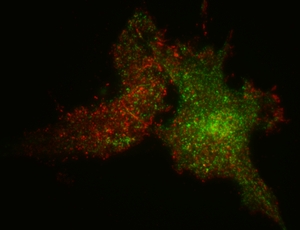
2. Visualization of receptor-induced signalling processes in living cells
Engineered
fusion proteins of
signalling proteins intramolecularly linked to various color variants
of the green fluorescent protein have fueled novel approaches to gather
information about the spatiotemporal orchestration of signal
transduction in living
cells. These genetically encoded fluorescent fusion proteins can be
directly visualised by
fluorescence microscopy without a need for additional cofactors. Thus,
they may serve to follow the localization of signaling proteins in
living
cells at the subcellular level and in a sub-second temporal resolution.
Signalling proteins that are being investigated in our group include
protein kinases C, phospholipases C, phosphatidylinositol-3-kinases,
receptor-tyrosine kinases, the nonreceptor tyrosine kinases Pyk2 and
Src and ion channels. Our scientific interest is to understand more
precisely the spatiotemporal properties and the interplay between
various signaling processes and their contribution to cell biological
responses.
Both methodological development and biological problems are addressed
in our group. Examples are i) the fingerprinting of spectrally
overlapping fluorochromes and its application to simultaneously record
multiple signalling processes in living cells, ii) fluorescence
resonance energy transfer (FRET) methods and their use in the
determination of ion channel assembly, protein interactions of
phosphoinositide-3-kinases and time-resolved monitoring of
intramlecular conformational changes, iii) quantitative fluorescence
recovery after photobleaching (FRAP) analysis to assess the lateral
mobility of membrane-associated or cytosolic signalling proteins and
iv) the quantitative analysis of ion concentrations and of the membrane
translocation during receptor-induced cell activation. To more
selectively observe signalling processes at the plasma membrane, we
have set up two fluorescence microscopes that operate in the total
internal reflection (TIR) mode. To obtain more information about the
protein function, the stability of protein-membrane interactions or
protein-protein interactions in the plasma membrane, these microscopes
can additionally measure FRET, FRAP, fluorescence lifetime or
anisotropy decay in the TIR
configuration.
Specific topics:
- Computer-assisted fingerprinting of signals emanating from spectrally
overlapping fluorochromes
- Receptor-induced
translocation of protein
kinases C
- Fluorescence resonance energy transfer FRET and various applications
- TIRF imaging technique
- Regulation and domain organization of phosphoinositide-3-kinase g
The group:
Dr. Kerstin Hill
(postdoc) group leader: Physiological and
pathophysiological roles of TRP channels
Isabelle
Straub (PhD student) topics: Pharmacological modulators of
Stim:Orai complexes, patch clamp analyses
Anke
Klein (PhD student) topics: Voltage sensor domains, protein
asymmetry in migrating cells
Melanie Kaiser (PhD student): Natural compounds modulate P2X7
Tanja Plötz (PhD student): Allosteric modulatory sites on P2X receptors
Beatrice
Oehler (MD student): Pharmacological modulation of
TRPA1
Cristoph Hempel (MD student): Novel modulators of P2X7
Marion
Leonhardt (technician) molecular biology, cell culture, lab
organisation
Nicole
Urban (technician) digital fluorescence video imaging,
screening, FLIPR
Helga
Sobottka (technician) cortical and hippocampal neurons,
calcium imaging, patch clamp
Former group members:
- (1995-1999) Thomas Hofmann (Dr. med., summa cum laude, Ernst-Reuter-Award of the FU Berlin, with Thomas Gudermann, now Emmy Noether stipend in Marburg)
- (2000-2004) Johannes Lenz (Schmiedeberg award of the DGPT)
- (2003-2008) Philipp Voigt (Dr. rer. nat., summa cum laude, now Leopoldina fellow in Danny Reinberg´s lab, NY)
- (2006-2008) Solveig Grossmann (Dr. rer. medic., summa cum laude, now with Enno Klußmann, FMP-Berlin)
- (2005-2008) Alejandra Pérez Sastre (Dr. rer. nat, magna cum laude, now Munich)
- (2001-2005) Daniel Sinnecker (Dr. med., summa cum laude, now Medical Faculty of the Technische Universität München)
- (2000-2002) Carsten Brock (Dr. rer. nat., magna cum laude, research fellow with Bernd Nürnberg, now Montpellier)
- (1999-2003) Nicole Hellwig (Dr. rer. nat., magna cum laude, now at the MDC-Berlin)
- (2003-2006) Umut Yilmaz (MD student)
- (2001-2003) Hemasse Amiri (MD student, now University of Heidelberg)
- (2006-2007) Kirstin Hobiger (biophysics diploma student)
- (2007-2008) Steffen Burgold (Photonics master student)
- (2007-2008) Marsha Wilke (biotechnology diploma student)
- (2004-2011) Astrid Tannert, post-doc biophysical imaging, protein:lipid interactions
Selected Publications:
Hill K, Schaefer M
(2008)
Ultraviolet
light and photosensitising agents activate TRPA1 via generation of
oxidative stress. Cell
Calcium (epub
ahead of print).
Tannert A, Voigt P,
Burgold S, Tannert S, Schaefer M
(2008)
Signal
amplification between Gbg release and PI3Kg-mediated
PI(3,4,5)P3 formation monitored by a fluorescent Gbg biosensor protein
and repetitive two component total internal reflection/fluorescence
redistribution after photobleaching analysis. Biochemistry (in
press).
Schauwienold D, Sastre
AP, Genzel N, Schaefer M, Reusch
HP (2008)
The
transactivated epidermal growth factor receptor recruits Pyk2 to
regulate Src kinase activity. J.
Biol. Chem.
283:27748-27756.
Pérez Sastre
A, Grossmann S, Reusch HP,
Schaefer M (2008)
Requirement
of an intermediate gene expression for biphasic ERK1/2 activation in
thrombin-stimulated vascular smooth muscle cells. J.
Biol. Chem.
283:25871-25878.
Hill K, Schaefer M
(2007)
TRPA1
is differentially modulated by the amphipathic molecules trinitrophenol
and chlorpromazine. J.
Biol. Chem.
282:7145-7153.
Wegierski T, Hill K,
Schaefer M, Walz G (2006)
The
HECT ubiquitin ligase AIP4 regulates the cell surface expression of
select TRP channels. EMBO
J. 25:5659-5669.
Voigt P, Dorner MB,
Schaefer M (2006)
Characterization
of p87PIKAP,
a novel regulatory subunit of
phosphoinositide 3-kinase g
that is highly expressed in heart and interacts with PDE3B. J.
Biol. Chem. 281:9977-9986.
Arniges M,
Fernandez-Fernandez JM, Albrecht N, Schaefer
M, Valverde MA (2006)
Human
TRPV4 channel splice variants revealed a key role of ankyrin domains in
multimerization and trafficking. J. Biol. Chem. 281:1580-1586.
Sinnecker D, Voigt P,
Hellwig N, Schaefer M (2005)
Reversible
photobleaching of enhanced green fluorescent proteins. Biochemistry
44:7085-7094.
Hellwig N, Albrecht N,
Harteneck C, Schultz G, Schaefer
M (2005)
Homo-
and heteromeric assembly of TRPV channel subunits.
J. Cell Sci. 118:917-928
Voigt P, Brock C,
Nürnberg B, Schaefer M (2004)
Assigning
functional domains within the p101 regulatory subunit of
phosphoinositide 3-kinase g.
J. Biol. Chem. 280:5121-5127.
Hellwig N, Plant TD,
Janson W, Schäfer M,
Schultz G, Schaefer M (2004)
TRPV1
acts as proton channel to induce acidification in nociceptive neurons.
J. Biol. Chem. 279:34553-34561.
Schaefer M, Mischak H,
Schnell S, Griese A, Iakubov R,
Riepenhausen G, Schöfl C (2004)
Mechanisms
of arginine-vasopressin-induced Ca2+
oscillations in beta-cells (HIT-T15): a role for oscillating protein
kinase C. Endocrinology 145:4635-4644.
Brock C, Schaefer M,
Reusch HP, Czupalla C, Michalke M,
Spicher K, Schultz G, Nürnberg B (2003)
Roles
of Gbg
in
membrane recruitment and activation of p110g/p101
phosphoinositide 3-kinase g.
J.
Cell Biol. 160:89-99.
Amiri H, Schultz G,
Schaefer M (2003)
FRET-based
analysis of TRPC subunit stoichiometry. Cell
Calcium 33:463-470.
Lenz JC, Reusch HP,
Albrecht N, Schultz G, Schaefer M
(2002)
Ca2+-controlled
competitive diacylglycerol binding of protein kinase C isoenzymes in
living cells. J.
Cell Biol. 159:291-302.
Hofmann T, Schaefer M,
Schultz G, Gudermann T. (2002)
Subunit
composition of mammalian transient receptor potential channels in
living cells. Proc.
Natl. Acad. Sci. USA 99:7461-7466.
Schaefer M, Albrecht N,
Hofmann T, Gudermann T, Schultz
G (2001)
Diffusion-limited
translocation mechanism of protein kinase C isotypes. FASEB
J. 15:1634-1636.
Schaefer M, Plant TD,
Stresow N, Albrecht N, Schultz G
(2002)
Functional
differences between TRPC4 splice variants. J. Biol.
Chem. 277:3752-3759.
Reusch HP, Schaefer M,
Plum C, Schultz G, Paul M (2001)
Gbg
mediate
differentiation of vascular smooth muscle cells. J.
Biol. Chem.276:19540-19547.
Schaefer M, Plant T,
Obukhov AG, Hofmann T, Gudermann T,
Schultz G (2000)
Receptor-mediated
regulation of the nonselective cation channels TRPC4 and TRPC5.
J.
Biol. Chem. 275:17517-17526.
Hofmann T, Schaefer M,
Schultz G, Gudermann T (2000)
Cloning,
expression and subcellular localization of two novel splice variants of
mouse transient receptor potential channel 2. Biochem.
J. 351:115-122.
Hofmann T, Obukhov AG,
Schaefer M, Harteneck C,
Gudermann T, Schultz G (1999)
Direct
activation of human TRPC6 and TRPC3 channels by diacylglycerol.
Nature 397:259-263.
Schaefer M, Hofmann T,
Schultz G, Gudermann T (1998)
A
new prostaglandin E receptor mediates calcium influx and acrosome
reaction in human spermatozoa. Proc.
Natl. Acad.
Sci. USA 95:3008-3013.
Patents:
Schaefer
M (1999) DE19915137
Quantifying
multiple fluorochromes in a sample comprises mathematical analysis of
fluorescence data spectrally resolved by using different excitation
wavelengths
Schaefer
M, Tannert A (2005) WO2007076839 / DE102005062673
Method
for determining a movement parameter for fluorochromes in a surrounding
medium
Publications
Michael Schaefer listed in PubMed