p_henri_research_01
Full size is 387 × 218 pixels
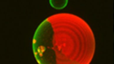
Example of giant unilamellar vesicles, phase-separated supported lipid bilayers and curved DNA origami nanostructures visualized using fluorescence confocal and atomic force microscopies. Image collected by Dr. Henri Franquelim, at Dep. Schwille, MPI Biochemistry.
Example of giant unilamellar vesicles, phase-separated supported lipid bilayers and curved DNA origami nanostructures visualized using fluorescence confocal and atomic force microscopies. Image collected by Dr. Henri Franquelim, at Dep. Schwille, MPI Biochemistry.