B11 – Die Rolle der EZM für die Knochenregeneration im Zebrafisch

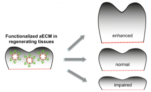
Der adulte Zebrafisch hat die bemerkenswerte Gabe, Organe und Gewebe nach schwerem Trauma vollständig zu regenerieren. Dies schließt die zügige Regeneration von Knochenstrukturen nach teilweisem Verlust der Flossen ein. Wir machen uns dieses Knochenregenerationsmodell zunutze, um den Einfluss der EZM auf die Knochenbildung zu studieren. Unser Ziel ist es, die Knochenbildung mit Hilfe artifizieller EZM zu steuern, insbesondere zu verbessern.

Publikationen

  1. Keil S, Gupta M, Brand M, Knopf F. Heparan sulfate proteoglycan expression in the regenerating zebrafish fin. Developmental Dynamics 1-13, 2021, doi.org/10.1002/dvdy.321.
  2. Dietrich K, Fiedler IA, Kurzyukova A, López-Delgado AC, McGowan LM, Geurtzen K, Hammond CL, Busse B, Knopf F. Skeletal biology and disease modeling in zebrafish. J Bone Miner Res 2021. doi: 10.1002/jbmr.4256 
  3. Schmidt JR*, Geurtzen K*, von Bergen M, Schubert K# and Knopf F#. Glucocorticoid treatment leads to aberrant ion and macromolecular transport in regenerating zebrafish fins. Front Endocrinol. 2019; 10:674. * equal contribution # co-corresponding authors.
  4. Geurtzen K, Knopf F. Adult Zebrafish Injury Models to Study the Effects of Prednisolone in Regenerating Bone Tissue. J Vis Exp. 2018; 140:e58429.
  5. Geurtzen K, Vernet A, Freidin A, Rauner M, Hofbauer LC, Schneider JE, Brand M, Knopf F. Immune Suppressive and Bone Inhibitory Effects of Prednisolone in Growing and Regenerating Zebrafish Tissues. J Bone Miner Res 2017, 32: 2476-2488.
  6. Geurtzen K*, Knopf F*, Wehner D, Huitema LF, Schulte-Merker S, Weidinger G. Mature steoblasts dedifferentiate in response to traumatic bone injury in the zebrafish fin and kull. Development. 2014;141:2225-34. (*geteilte Erstautorschaft).
  7. Gupta M, Brand M. Identification and Expression Analysis of Zebrafish Glypicans during Embryonic Development. PLoS One. 2013;8:e80824.
    Boekel C, Brand M. Generation and interpretation of FGF morphogen gradients in vertebrates. Curr Opin Gen Dev. 2013;23:415-22.
  8. Knopf F, Hammond C, Chekuru A, Kurth T, Hans S, Weber CW, Mahatma G, Fisher S, Brand M, Schulte-Merker S, Weidinger G. Bone regenerates via dedifferentiation of osteoblastsen the zebrafish fin. Dev Cell. 2011;20:713-24.
  9. Nowak M, Yu SR, Gupta M, Machate A, Brand M. Interpretation of the FGF8 morphogen gradient is regulated by endocytic trafficking. Nat Cell Biol. 2011;13:153-8.
  10. Yu SR, Burkhardt M, Nowak M, Ries J, Petrášek Z, Scholpp S, Schwille P*, Brand M.* FGF8 morphogen gradient is formed by a source-sink mechanism with freely-diffusing molecules. Nature. 2009;461:533-6. (*geteilte Seniorautorschaft).
  11. Ries J, Yu SR, Burkhardt M, Brand M*, Schwille P*. Modular scanning FCS quantifies ligand-receptor interactions in live multicellular organisms. Nat Methods. 2009;6:643-5. (*geteilte Seniorautorschaft).
  12. Raible F, Brand M. Divide et Impera-the midbrain-hindbrain boundary and its organizer. Trends Neurosci. 2004;27:727-34.
    Scholpp S, Brand M. Endocytosis controls spreading and effective signaling range of FGF8. Curr Biol. 2004;14:1834-41.

Kontakt

Prof. Dr. Michael Brand

Technische Universität Dresden
CRTD – Zentrum für Regenerative Therapien
Fetscherstraße 105, 01307 Dresden

Telefon: +49 (0)351 458 82300
E-Mail: michael.brand@tu-dresden.de
Web: crt-dresden.de/research/research-groups

Jun.-Prof. Dr. Franziska Knopf

Technische Universität Dresden
CRTD – Zentrum für Regenerative Therapien TU Dresden
Fetscherstraße 105, 01307 Dresden

Telefon: +49 (0)351 458 82336
E-Mail: franziska.knopf@tu-dresden.de
Web: crt-dresden.de/research/research-groups