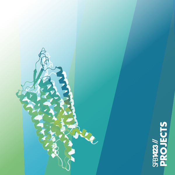

Project A05 – Structures of adhesion GPCR by cryo-electron microscopy
Several 3D-structures of adhesion GPCRs (aGPCRs) have been solved, but this information is still limited to a few receptor examples, partial structures and mostly active conformations of the transmembrane domain (TMD). An important open question is the interplay between the TMD and the N terminus. The N terminus, with up to 6.000 amino acids, is predisposed to integrate a variety of signals such as ligand binding, cell-cell (matrix) contacts and even mechanical forces into intracellular signals. Various important general and detailed steps in this signaling process are not yet understood, partly due to a lack of structural information. Therefore, project A05 aims to study the 3D-structures of different full-length aGPCRs and aGPCR complexes, either in (in-)active or basal conformation, using single-particle cryo-electron microscopy and cryo-electron tomography.
In contrast to most other GPCRs, adhesion GPCRs (aGPCRs) exhibit very large N-termini (up to 6,000 amino acids) constituted by structurally and functionally diverse domains. These aGPCRs bind to a number of different proteins or to other extracellular interaction partners. The N-termini, therefore, are predestined to integrate a multitude of signals, such as ligand binding, cell-cell (-matrix) contacts and even mechanical forces finally into intracellular signals. The project A05 aims to solve 3D-structures of aGPCRs by cryo-EM (or protein X-ray crystallography for receptor parts) to unravel the activation mechanisms of this unique class of membrane receptors.
Contact

Dr. Patrick Scheerer (Project Leader)
Charité - Universitätsmedizin Berlin
Institute for Medical Physics and Biophysics
Group Structural Biology of Cellular Signaling
(Protein X-ray Crystallography - Cryo-Electron Microscopy - Signal Transduction)
Charitéplatz 1, D-10117 Berlin
Phone +49 30 450 524 178
E-Mail
Web biophysik.charite.de/forschung/ag_scheerer

Prof. Dr. Christian M.T. Spahn (Project Leader)
Charité - Universitätsmedizin Berlin
Institute for Medical Physics and Biophysics
AG Cryo-electron microscopy of macromolecular machines
Charitéplatz 1, D-10117 Berlin
Phone +49 30 450 524 131
E-Mail
Web biophysik.charite.de/forschung/ag_spahn

Prof. Dr. Torsten Schöneberg (Project Leader)
Leipzig University, Faculty of Medicine
Rudolf Schönheimer Institute of Biochemistry
Johannisallee 30, D-04103 Leipzig
Phone +49 341 97 22150
E-Mail
Web uniklinikum-leipzig.de/ag-schöneberg

Dr. Amal Hassan (Postdoctoral Fellow)
Charité - Universitätsmedizin Berlin
Institute for Medical Physics and Biophysics
Group Structural Biology of Cellular Signaling
(Protein X-ray Crystallography - Cryo-Electron Microscopy - Signal Transduction)
Charitéplatz 1, D-10117 Berlin
Phone: +49 30 450 524 150
E-Mail:

Franziska Wiechert (Phd Student)
Charité - Universitätsmedizin Berlin
Institute for Medical Physics and Biophysics
Group Structural Biology of Cellular Signaling
(Protein X-ray Crystallography - Cryo-Electron Microscopy - Signal Transduction)
Charitéplatz 1, D-10117 Berlin
Phone: +49 30 450 524 135
E-Mail:

Further Team Members
Dr. Gunnar Kleinau (Senior Scientist at Charité - Group Scheerer)
Dr. Michal Szczepek (A01 - Group Scheerer)
Dr. Andrea Schmidt (Senior scientist at Charité - Group Scheerer)
David Speck (PhD student A05/A01 - Group Scheerer)
Anja Koch (Technician at Charité - Group Scheerer)
Justus Loerke (Senior Scientist at Charité - Group Spahn)
Magdalena Schacherl (Senior Scientist at Charité - Group Spahn)
Jörg Bürger (Technician at Charité - Group Spahn)
Resources
Methods and Techniques used in A05 at the Charité:
Structure biology methods:
- Protein X-ray crystallography, membrane protein crystallization, lipidic cubic phase (LCP) crystallization (e.g. TTP Labtech’s mosquito, Gryphon-LCP crystallization and PRIMA Xtallob robots; Formulatrix – Rock Imager 182/54; High-end Stereo microscope Leica M205; Formulatrix – MUVIS)
- X-ray data acquisition: synchrotrons (e.g. BESSY and DESY (Germany), ESRF (France), or in house rotating anode generator (MicroMax007 Microfocus)
- Cryo-electron tomography (cryo-ET) setup – cryo-FIBSEM dual beam instrument (ThermoFisher Aquilos2) and a confocal cryo-LM (LEICA SP8)
- Cryo-electron microscopy (in house) – high-end 300 kV FEI Titan Krios G3 cyro-TEM System/ K3 direct electron detector/ Volta phase plate/ energy filter, Vitrobot, 120kV TEM for sample screening
GPCR production methods and signaling/ligand-binding assays:
- GPCR cloning/expression/purification/solubilisation methods
- Large-scale heterologous cell expression (baculovirus expression in Sf9/High Five™ and HEK or Expi293F and E.Coli)
- Various purification systems: Äktapurifier (FPLC), AktaprimePlus (Gel filtration) or ultra-high-performance liquid chromatography – UltiMate 3000 Bio UHPLC
- Production of G-protein or arrestin
- Nanodisc/SMA lipid particles (SMALPs) production and integration
- NanoBRET Ligand-binding and G-protein dissociation assays
- cAMP signaling assays (in vivo)
Biophysical methods:
- Nano-differential scanning fluorimetry (nDSF with NanoTemper Prometheus system) or other thermal shift assays (e.g. CPM) to test thermostability
- MicroScale Thermophoresis (MST) to test binding affinity & protein interactions (NanoTemper Monolith NT 115 system)
- Multiplate reader for assay development with e.g. BRET/FRET/nanoLuc (CLARIOstar Plus – BMG Labtech)
- Surface plasmon resonance-like instrument to test biomolecular interaction (White FOx 1.0 – FOx Biosystem)
- Static light scattering – Multi-angle light scattering detector (Wyatt Technology)
- UV-vis spectrometer (Cary4000, Agilent)
Publications
Batebi H, Pérez-Hernández G, Rahman SN, Lan B, Kamprad K, Shi M, Speck D, Tiemann JKS, Guixà-González R, Reinhardt F, Stadler PF, Papasergi-Scott MM, Skiniotis G, Scheerer P, Kobilka BK, Mathiesen JM, Liu X, Hildebrand PW. Mechanistic insights into G-protein coupling with an agonist-bound G-protein-coupled receptor. Nature Struct Mol Biol (2024). https://doi.org/10.1038/s41594-024-01334-2 .
Makkonen K, Jännäri M, Crisóstomo L, Kuusi M, Patyra K, Melnyk V, Linnossuo V, Ojala J, Ravi R, Löf C, Mäkelä JA, Miettinen P, Laakso S, Ojaniemi M, Jääskeläinen J, Laakso M, Bossowski F, Sawicka B, Stożek K, Bossowski A, Kleinau G, Scheerer P, FinnGen F, Reeve MP, Kero J. Mechanisms of thyrotropin receptor-mediated phenotype variability deciphered by gene mutations and M453T-knockin model. JCI Insight. 2024 Jan 9;9(4):e167092. doi: 10.1172/jci.insight.167092 . PMID: 38194289 .
Entsie P, Kang Y, Amoafo EB, Schöneberg T, Liverani E. The Signaling Pathway of the ADP Receptor P2Y12 in the Immune System: Recent Discoveries and New Challenges. Int J Mol Sci. 2023; 24:6709.
Frenster JD, Erdjument-Bromage H, Stephan G, Ravn-Boess N, Wang S, Liu W, Bready D, Wilcox J, Kieslich B, Jankovic M, Wilde C, Horn S, Sträter N, Liebscher I, Schöneberg T, Fenyo D, Neubert TA, Placantonakis DG. PTK7 is a positive allosteric modulator of GPR133 signaling in glioblastoma. Cell Rep. 2023 Jun 23;42(7):112679.
Frenster JD, Erdjument-Bromage H, Liu W, Stephan G, Ravn-Boess N, Bready D, Wilcox J, Kieslich B, Jankovic M, Wilde C, Horn S, Sträter N, Liebscher I, Schöneberg T, Fenyo D, Neubert TA, Placantonakis DG. PTK7 is a positive allosteric modulator of GPR133 (ADGRD1) signaling in GBM. biorxiv. 2022.06. 15.496232.
Kleinau G, Ali AH, Wiechert F, Szczepek M, Schmidt A, Spahn CMT, Liebscher I, Schöneberg T, Scheerer P. Intramolecular activity regulation of adhesion GPCRs in light of recent structural and evolutionary information. Pharmacol Res. 2023 Nov;197:106971. doi: 10.1016/j.phrs.2023.106971. Epub 2023 Oct 30. PMID: 38032292.
Groß VE, Gershkovich MM, Schöneberg T, Kaiser A, Prömel S. NanoBRET in C. elegans illuminates functional receptor interactions in real time. BMC Mol Cell Biol. 2022; 23(1):8. DOI: 10.1186/s12860-022-00405-w.
Liebscher I, Schöneberg T, Thor D. Stachel-mediated activation of adhesion G protein-coupled receptors: insights from cryo-EM studies. Signal Transduct Target Ther. 2022; 7:227.
Matúš D, Post WB, Horn S, Schöneberg T, Prömel S. Latrophilin-1 drives neuron morphogenesis and shapes chemo- and mechanosensation-dependent behavior in C. elegans via a trans function. Biochem Biophys Res Commun. 2022; 589:152-158. DOI: 10.1016/j.bbrc.2021.12.006.
Philippe A, Kleinau G, Gruner JJ, Wu S, Postpieszala D, Speck D, Heidecke H, Dowell SJ, Riemekasten G, Hildebrand PW, Kamhieh-Milz J, Catar R, Szczepek M, Dragun D, Scheerer P. Molecular Effects of Auto-Antibodies on Angiotensin II Type 1 Receptor Signaling and Cell Proliferation. Int J Mol Sci. 2022 Apr 2;23(7):3984. doi: 10.3390/ijms23073984. PMID: 35409344
Ping YQ, Xiao P, Yang F, Zhao RJ, Guo SC, Yan X, Wu X, Zhang C, Lu Y, Zhao F, Zhou F, Xi YT, Yin W, Liu FZ, He DF, Zhang DL, Zhu ZL, Jiang Y, Du L, Feng SQ, Schöneberg T, Liebscher I, Xu HE, Sun JP. Structural basis for the tethered peptide activation of adhesion GPCRs. Nature. 2022. https://doi.org/10.1038/s41586-022-04619-y.
Schulze AS, Kleinau G, Krakowsky R, Rochmann D, Das R, Worth CL, Krumbholz P, Scheerer P, Stäubert C. Evolutionary analyses reveal immune cell receptor GPR84 as a conserved receptor for bacteria-derived molecules. iScience. 2022; 25:105087.
Speck D, Kleinau G, Meininghaus M, Erbe A, Einfeldt A, Szczepek M, Scheerer P, Pütter V. Expression and Characterization of Relaxin Family Peptide Receptor 1 Variants. Front Pharmacol. 2022 Jan 28;12:826112. doi: 10.3389/fphar.2021.826112. eCollection 2021. PMID: 35153771 Free PMC article.
Wilde C, Mitgau J, Suchý T, Schoeneberg T, Liebscher I. Translating the Force – mechano-sensing GPCRs. Am J Physiol Cell Physiol. 2022 Apr 13. doi: 10.1152/ajpcell.00465.2021.PMID: 35417266 Review.
Yang Y, Stensitzki T, Sauthof L, Schmidt A, Piwowarski P, Velazquez Escobar F, Michael N, Nguyen AD, Szczepek M, Brünig FN, Netz RR, Mroginski MA, Adam S, Bartl F, Schapiro I, Hildebrandt P, Scheerer P, Heyne K. Ultrafast proton-coupled isomerization in the phototransformation of phytochrome. Nat Chem. 2022 May 16. doi: 10.1038/s41557-022-00944-x. PMID: 35577919.
Zimmermann A, Vu O, Brüser A, Sliwoski G, Marnett LJ, Meiler J, Schöneberg T. Mapping the Binding Sites of UDP and Prostaglandin E2 Glyceryl Ester in the Nucleotide Receptor P2Y6. ChemMedChem. 2022, e202100683.
Catar R, Herse-Naether M, Zhu N, Wagner P, Wischnewski O, Kusch A, Kamhieh-Milz J, Eisenreich A, Rauch U, Hegner B, Heidecke H, Kill A, Riemekasten G, Kleinau G, Scheerer P, Dragun D, Philippe A. Autoantibodies Targeting AT1- and ETA-Receptors Link Endothelial Proliferation and Coagulation via Ets-1 Transcription Factor. Int J Mol Sci. 2022, 23, 244. doi: 10.3390/ijms23010244. PMID: 35008670.
Frenster JD, Stephan G, Ravn-Boess N, Bready D, Wilcox J, Kieslich B, Wilde C, Sträter N, Wiggin GR, Liebscher I, Schöneberg T, Placantonakis DG. Functional impact of intramolecular cleavage and dissociation of adhesion G protein-coupled receptor GPR133 (ADGRD1) on canonical signaling. J Biol Chem. 2021; 296:100798. DOI: 10.1016/j.jbc.2021.100798.
Heyder NA, Kleinau G, Speck D, Schmidt A, Paisdzior S, Szczepek M, Bauer B, Koch A, Gallandi M, Kwiatkowski D, Bürger J, Mielke T, Beck-Sickinger A, Hildebrand P, Spahn CMT, Hilger D, Schacherl M, Biebermann H, Hilal T, Kühnen P, Kobilka BK, Scheerer P. Structures of active melanocortin-4 receptor−Gs-protein complexes with NDP-α-MSH and setmelanotide. Cell Research. 2021, 31(11):1176-1189. doi: 10.1038/s41422-021-00569-8. PMID: 34561620.
Kaczmarek I, Suchý T, Prömel S, Schöneberg T, Liebscher I, Thor D. The relevance of adhesion G protein-coupled receptors in metabolic functions. Biol Chem. 2021; 403(2):195-209. doi: 10.1515/hsz-2021-0146.
Schöneberg T, Liebscher I. Mutations in G Protein–Coupled Receptors: Mechanisms, Pathophysiology and Potential Therapeutic Approaches. Pharamcological Reviews. 2021; 73:89-119. doi: https://doi.org/10.1124/pharmrev.120.000011.
Wallach T, Mossmann ZJ, Szczepek M, Wetzel M, Machado R, Raden M, Miladi M, Kleinau G, Krüger C, Dembny P, Adler D, Zhai Y, Kumbol V, Dzaye O, Schüler J, Futschik M, Backofen R, Scheerer P, Lehnardt S. MicroRNA-100-5p and microRNA-298-5p released from apoptotic cortical neurons are endogenous Toll-like receptor 7/8 ligands that contribute to neurodegeneration. Mol Neurodegener. 2021 Nov 27;16(1):80. doi: 10.1186/s13024-021-00498-5.
Wittlake A, Prömel S, Schöneberg T. The Evolutionary History of Vertebrate Adhesion GPCRs and Its Implication on Their Classification. Int J Mol Sci. 2021; 22(21):11803. DOI: 10.3390/ijms222111803.
Frenster JD, Kader M, Kamen S, Sun J, Chiriboga L, Serrano J, Bready D, Golub D, Ravn-Boess N, Stephan G, Chi AS, Kurz SC, Jain R, Park CY, Fenyo D, Liebscher I, Schöneberg T, Wiggin G, Newman R, Barnes M, Dickson JK, MacNeil DJ, Huang X, Shohdy N, Snuderl M, Zagzag D, Placantonakis DG. Expression profiling of the adhesion G protein-coupled receptor GPR133 (ADGRD1) in glioma subtypes. Neurooncol Adv. 2020; 2(1):vdaa053. DOI: 10.1093/noajnl/vdaa053.
Kleinau G, Heyder NA, Tao YX, Scheerer P. Structural Complexity and Plasticity of Signaling Regulation at the Melanocortin-4 Receptor. Int J Mol Sci. 2020 Aug 10;21(16):5728. doi: 10.3390/ijms21165728. PMID: 32785054
Paisdzior S, Dimitriou IM, Schöpe PC, Annibale P, Scheerer P, Krude H, Lohse MJ, Biebermann H, Kühnen P. Differential Signaling Profiles of MC4R Mutations with Three Different Ligands. Int J Mol Sci. 2020 Feb 12;21(4). pii: E1224. doi: 10.3390/ijms21041224. PMID: 32059383
Schulze A, Kleinau G, Neumann S, Scheerer P, Schöneberg T, Brüser A. The intramolecular agonist is obligate for activation of glycoprotein hormone receptors. FASEB J. 2020 Jul 10. doi: 10.1096/fj.202000100R. Epub ahead of print. PMID: 32648604
Hamann J, Aust G, Araç D, Engel FB, Formstone C, Fredriksson R, Hall RA, Harty BL, Kirchhoff C, Knapp B, Krishnan A, Liebscher I, Lin HH, Martinelli DC, Monk KR, Peeters MC, Piao X, Prömel S, Schöneberg T, Schwartz TW, Singer K, Stacey M, Ushkaryov YA, Vallon M, Wolfrum U, Wright MW, Xu L, Langenhan T, Schiöth HB. A tethered agonist within the ectodomain activates the adhesion G protein-coupled receptors GPR126 and GPR133. Pharmacol Rev. 2015; 67:338-67.
Bohnekamp J, Schöneberg T. Cell adhesion receptor GPR133 couples to Gs protein. J Biol Chem. 2011; 286:41912-6.
Liebscher I, Schön J, Petersen SC, Fischer L, Auerbach N, Demberg LM, Mogha A, Cöster M, Simon KU, Rothemund S, Monk KR, Schöneberg T. A tethered agonist within the ectodomain activates the adhesion G protein-coupled receptors GPR126 and GPR133. Cell Rep. 2014; 9:2018-26.
Schöneberg T, Kleinau G, Brüser A. What are they waiting for? – Tethered agonism in G protein-coupled receptors. Pharmacol Res. 2016; 108:9-15.
Prömel S, Frickenhaus M, Hughes S, Mestek L, Staunton D, Woollard A, Vakonakis I, Schöneberg T, Schnabel R, Russ AP, Langenhan T. The GPS motif is a molecular switch for bimodal activities of adhesion class G protein-coupled receptors. Cell Rep. 2012; 2:321-31.
Behrmann E, Loerke J, Budkevich TV, Yamamoto K, Schmidt A, Penczek PA, Vos MR, Bürger J, Mielke T, Scheerer P, Spahn CM. Structural snapshots of actively translating human ribosomes. Cell. 2015; 161:845-57.
Yamamoto H, Collier M, Loerke J, Ismer J, Schmidt A, Hilal T, Sprink T, Yamamoto K, Mielke T, Bürger J, Shaikh TR, Dabrowski M, Hildebrand PW, Scheerer P, Spahn CM. Molecular architecture of the ribosome-bound Hepatitis C Virus internal ribosomal entry site RNA. EMBO J. 2015; 34:3042-58.
Nikolay R, Hilal T, Qin B, Mielke T, Bürger J, Loerke J, Textoris-Taube K, Nierhaus KH, Spahn CMT. Structural Visualization of the Formation and Activation of the 50S Ribosomal Subunit during In vitro. Mol Cell. 2018; 70:881-93.
Szczepek M, Beyrière F, Hofmann KP, Elgeti M, Kazmin R, Rose A, Bartl FJ, von Stetten D, Heck M, Sommer ME, Hildebrand PW, Scheerer P. Crystal structure of a common GPCR-binding interface for G protein and arrestin. Nature Commun. 2014; 5:4801.
Qureshi BM, Schmidt A, Behrmann E, Bürger J, Mielke T, Spahn CMT, Heck M, Scheerer P. Mechanistic insights into the role of prenyl-binding protein PrBP/δ in membrane dissociation of phosphodiesterase 6. Nature Commun. 2018; 9:90.
